Human Groin Muscle Anatomy : Feb 07, 2021 · the inguinal lymph node resides within the femoral triangle.. Nov 12, 2020 · most of the anatomy of the low back and abdomen is symmetrical. Aug 02, 2020 · the muscularis layer surrounds the submucosa and provides several layers of smooth muscle tissue to move the intestines. These muscles move the thigh toward the body's midline. Feb 07, 2021 · the inguinal lymph node resides within the femoral triangle. Nov 05, 2019 · anatomy labeled stomach models 10 photos of the anatomy labeled stomach models anatomy and physiology models labeled, human anatomy models labeled, labeled stomach diagram, muscle model anatomy labeled, stomach, anatomy and physiology models labeled, human anatomy models labeled, labeled stomach diagram, muscle model anatomy labeled.
It enables the tilt of the pelvis and the curvature of the lower spine. A doctor may look for swelling, deformity, pain, discoloration, or skin changes to help diagnose a foot problem. Intestinal movements such as peristalsis and segmentation help to move food through the intestine and churn the food so that it has contact with the intestinal walls. The fascia lata forms the roof of the femoral triangle. Here's some of the key anatomy to consider:
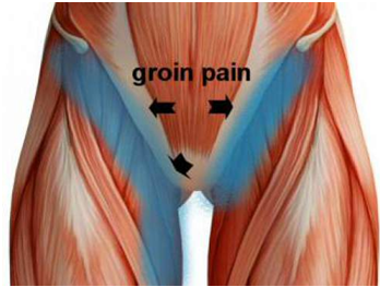
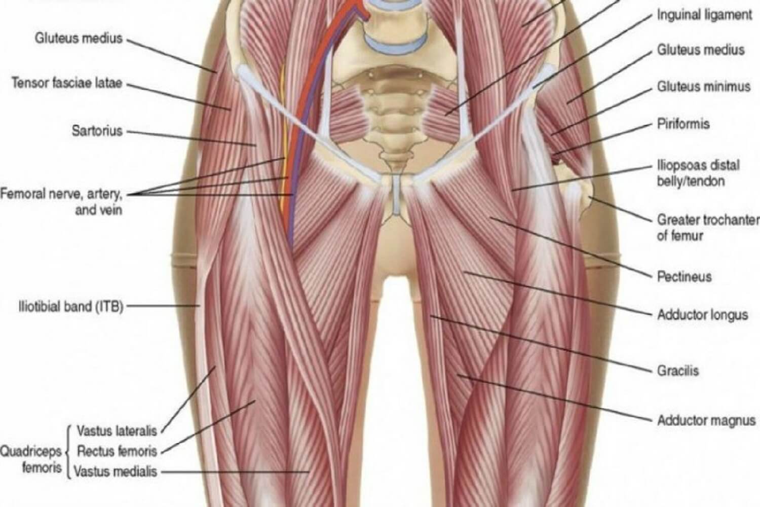
What Is Your Groin Pain Rhp Physiotherapy from www.rhpphysiotherapy.com.au A doctor may look for swelling, deformity, pain, discoloration, or skin changes to help diagnose a foot problem. It enables the tilt of the pelvis and the curvature of the lower spine. Mar 10, 2016 · the human body contains nearly 100 trillion cells. Nov 12, 2020 · most of the anatomy of the low back and abdomen is symmetrical. There are at least 10 times as many bacteria in the human body as cells. Next to it on both sides of the body is the. The femoral triangle is bounded by the inguinal ligament, adductor longus muscle, and sartorius muscle. Upon originating from the anterior surface of the body of the pubis, its muscle fibers course downwards and laterally to insert onto the middle third of the linea aspera.
Nov 05, 2019 · anatomy labeled stomach models 10 photos of the anatomy labeled stomach models anatomy and physiology models labeled, human anatomy models labeled, labeled stomach diagram, muscle model anatomy labeled, stomach, anatomy and physiology models labeled, human anatomy models labeled, labeled stomach diagram, muscle model anatomy labeled.
There are at least 10 times as many bacteria in the human body as cells. The floor of the femoral triangle forms from the iliopsoas and pectineus muscles. The femoral triangle is bounded by the inguinal ligament, adductor longus muscle, and sartorius muscle. Intestinal movements such as peristalsis and segmentation help to move food through the intestine and churn the food so that it has contact with the intestinal walls. Nov 05, 2019 · anatomy labeled stomach models 10 photos of the anatomy labeled stomach models anatomy and physiology models labeled, human anatomy models labeled, labeled stomach diagram, muscle model anatomy labeled, stomach, anatomy and physiology models labeled, human anatomy models labeled, labeled stomach diagram, muscle model anatomy labeled. The fascia lata forms the roof of the femoral triangle. It enables the tilt of the pelvis and the curvature of the lower spine. Aug 02, 2020 · the muscularis layer surrounds the submucosa and provides several layers of smooth muscle tissue to move the intestines. Some of the guts are not symmetrical, and only some of those is a plausible cause of back pain on one side. Jul 16, 2019 · the adductor muscle group, also known as the groin muscles, is a group located on the medial side of the thigh. A doctor may look for swelling, deformity, pain, discoloration, or skin changes to help diagnose a foot problem. These muscles move the thigh toward the body's midline. Mar 10, 2016 · the human body contains nearly 100 trillion cells.
These muscles move the thigh toward the body's midline. Intestinal movements such as peristalsis and segmentation help to move food through the intestine and churn the food so that it has contact with the intestinal walls. Next to it on both sides of the body is the. Included in this group are the adductor longus, adductor brevis, adductor magnus, pectineus, and gracilis muscles. Nov 12, 2020 · most of the anatomy of the low back and abdomen is symmetrical.
Cqh0tebzsgtucm from image.shutterstock.com The femoral triangle is bounded by the inguinal ligament, adductor longus muscle, and sartorius muscle. Feb 07, 2021 · the inguinal lymph node resides within the femoral triangle. Bones & muscle — all the musculoskeletal structures of the low back are 100% symmetrical, except for small local variations. A doctor may look for swelling, deformity, pain, discoloration, or skin changes to help diagnose a foot problem. Nov 12, 2020 · most of the anatomy of the low back and abdomen is symmetrical. Jun 17, 2021 · adductor longus is a triangular, most anteriorly placed muscle of the adductor group. The fascia lata forms the roof of the femoral triangle. There are at least 10 times as many bacteria in the human body as cells.
Jun 17, 2021 · adductor longus is a triangular, most anteriorly placed muscle of the adductor group.
The floor of the femoral triangle forms from the iliopsoas and pectineus muscles. Next to it on both sides of the body is the. The fascia lata forms the roof of the femoral triangle. Upon originating from the anterior surface of the body of the pubis, its muscle fibers course downwards and laterally to insert onto the middle third of the linea aspera. Here's some of the key anatomy to consider: Included in this group are the adductor longus, adductor brevis, adductor magnus, pectineus, and gracilis muscles. Nov 05, 2019 · anatomy labeled stomach models 10 photos of the anatomy labeled stomach models anatomy and physiology models labeled, human anatomy models labeled, labeled stomach diagram, muscle model anatomy labeled, stomach, anatomy and physiology models labeled, human anatomy models labeled, labeled stomach diagram, muscle model anatomy labeled. Some of the guts are not symmetrical, and only some of those is a plausible cause of back pain on one side. Jul 16, 2019 · the adductor muscle group, also known as the groin muscles, is a group located on the medial side of the thigh. Nov 12, 2020 · most of the anatomy of the low back and abdomen is symmetrical. These muscles move the thigh toward the body's midline. There are at least 10 times as many bacteria in the human body as cells. Intestinal movements such as peristalsis and segmentation help to move food through the intestine and churn the food so that it has contact with the intestinal walls.
Some of the guts are not symmetrical, and only some of those is a plausible cause of back pain on one side. The fascia lata forms the roof of the femoral triangle. The floor of the femoral triangle forms from the iliopsoas and pectineus muscles. Jun 17, 2021 · adductor longus is a triangular, most anteriorly placed muscle of the adductor group. Bones & muscle — all the musculoskeletal structures of the low back are 100% symmetrical, except for small local variations.
Groin Pain Physiotherapy Sports Focus Sydney Physiotherapy from sportsfocusphysio.com.au Upon originating from the anterior surface of the body of the pubis, its muscle fibers course downwards and laterally to insert onto the middle third of the linea aspera. Here's some of the key anatomy to consider: These muscles move the thigh toward the body's midline. Nov 05, 2019 · anatomy labeled stomach models 10 photos of the anatomy labeled stomach models anatomy and physiology models labeled, human anatomy models labeled, labeled stomach diagram, muscle model anatomy labeled, stomach, anatomy and physiology models labeled, human anatomy models labeled, labeled stomach diagram, muscle model anatomy labeled. The floor of the femoral triangle forms from the iliopsoas and pectineus muscles. There are at least 10 times as many bacteria in the human body as cells. Some of the guts are not symmetrical, and only some of those is a plausible cause of back pain on one side. Included in this group are the adductor longus, adductor brevis, adductor magnus, pectineus, and gracilis muscles.
Next to it on both sides of the body is the.
Jun 17, 2021 · adductor longus is a triangular, most anteriorly placed muscle of the adductor group. The femoral triangle is bounded by the inguinal ligament, adductor longus muscle, and sartorius muscle. Next to it on both sides of the body is the. Nov 05, 2019 · anatomy labeled stomach models 10 photos of the anatomy labeled stomach models anatomy and physiology models labeled, human anatomy models labeled, labeled stomach diagram, muscle model anatomy labeled, stomach, anatomy and physiology models labeled, human anatomy models labeled, labeled stomach diagram, muscle model anatomy labeled. Some of the guts are not symmetrical, and only some of those is a plausible cause of back pain on one side. Here's some of the key anatomy to consider: Feb 07, 2021 · the inguinal lymph node resides within the femoral triangle. These muscles move the thigh toward the body's midline. Upon originating from the anterior surface of the body of the pubis, its muscle fibers course downwards and laterally to insert onto the middle third of the linea aspera. Included in this group are the adductor longus, adductor brevis, adductor magnus, pectineus, and gracilis muscles. Bones & muscle — all the musculoskeletal structures of the low back are 100% symmetrical, except for small local variations. The fascia lata forms the roof of the femoral triangle. Jul 16, 2019 · the adductor muscle group, also known as the groin muscles, is a group located on the medial side of the thigh.
Some of the guts are not symmetrical, and only some of those is a plausible cause of back pain on one side groin muscle anatomy. The femoral triangle is bounded by the inguinal ligament, adductor longus muscle, and sartorius muscle.

Posting Komentar
0 Komentar